Thursday, January 05, 2006

SNMI 2005 meeting report


I attended the 37th Annual Conference of Society of Nuclear Medicine, India (SNMI) 2005 held at Calicut in the state of Kerala, India from 7th -11th December, 2005. It was the first time for me to attend an International conference related to Nuclear medicine held in India.
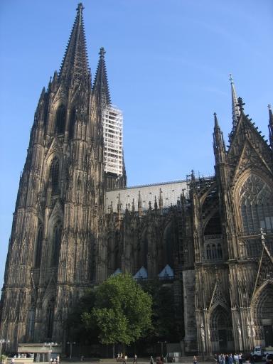
The conference venue was at Kadavu Resort, located in a sprawling nine-acre plot along the side of the backwaters, with groves of swaying, graceful coconut palms facing the dancing ripples on the water.
The theme of the conference was chosen to be “Scroll into the era of Fusion Imaging” to reflect the rapid changes and the future opportunities of Nuclear Medicine in India with a global perspective.
The Scientific Programme consisted of Keynote addresses, plenary lectures and Specialty sessions delivered by pioneers and experts of Nuclear medicine and molecular imaging from India and abroad. In addition, symposia, panel discussion and free paper sessions were also part of the programme. There were nearly 250 delegates including students, with experts coming from USA, UK, Singapore, Hong Kong and Korea. I was the only representative from Japan. There were 35 poster presentations and 40 oral presentations in free paper session covering various clinical and basic research topics of Nuclear medicine.
The welcome address was given by Dr. P.S. Soni, Head, PET Cyclotron Division, Radiation Medicine Centre, Mumbai, India. The initial day’s lectures had two sessions - Fusion Imaging (Technical and Clinical) and Gastroenterology coupled with symposium on “sentinel node biopsy” and a panel discussion on “Interstitial lung disease”. The fusion imaging lectures covered basics and radiation safety of SPECT-CT and PET-CT fusion imaging. The significant factors contributing to attenuation correction accuracy of fused images such as CT acquisition time, motion artifacts, slice thickness variations and reconstruction strategies were mentioned. There were also talks on clinical usage of SPECT-CT in skeletal, infectious and oncological disorders. Few clinical cases of implant infections and pyrexia of unknown origin (PUO) were imaged with 99mTc-labelled leukocyte or Gallium SPECT-CT and accurate localization and quantification of the infection was reported. In the Gastroenterology session, talks were given on the relative challenges of ERCP and MRCP in hepatobiliary scintigraphy. Advances in the usage of radioisotope liver scan for diffuse hepatic disease was also presented in the same session.

In the next day among the special orations, Homi Bhaba oration was given by Dr Joseph Mantil, who gave a general talk of tools of Molecular Imaging currently available and stressed on importance of early diagnosis of atherosclerosis using FDG imaging. In the cardiology session, benefits of myocardial perfusion scan were discussed. The imaging techniques other than conventional coronary angiography in patient management such as quantifying atheroma burden using Multislice CT coronary calcium scoring or EBCT, use of perfusion scan as prognostic indicator of cardiac dysfunction, use of perfusion MR imaging to localize subendocardial hypoperfusion were presented. Among the free paper session, few lectures were of notable interest. Most of them were either clinical studies or evaluation of cyclotron or SPECT-CT imaging applications. An indigenous user friendly and easy to use Quality control test kit for 99mTc-radiopharmaceuticals called QTECH was introduced. My talk was scheduled in the post-lunch session and as expected it gathered little audience after heavy meals! Though there were not many queries immediately after my talk, a few came up during tea break.

The next day’s session was entirely devoted to applications of PET-CT in different clinical disorders such as Lymphoma, breast cancer, thyroid cancer and other benign disorders. Here I would like to mention that currently there are only two functional cyclotrons in the country – one at Mumbai (BARC) and other at Hyderabad (Apollo Hospital). There is only one dedicated PET camera at Tata Memorial Hospital and three coincidence gamma PET cameras at Hinduja Hospital, Jaslok Hospital and Bombay Hospital all being in Mumbai. At Hyderabad, a PET-CT machine was installed this June and has started functioning. Squarely speaking, this is a negligible contribution to a country having a population of more than a billion and they too are recent installations. The major reasons for this include cost-effectiveness, government restrictions and low awareness among the health care planners and medical fraternity outside imaging community. But with increasing pressure from imaging societies and multinational companies, over the coming 2-3 years, plans have been charted to install few more cyclotrons and PET-CT in Delhi, Kolkata and Bangalore. The need for them was well appraised with leading nuclear medicine specialists giving talks on forthcoming clinical applications of PET-CT.
The final day was concluded with plenary lecture from Dr. June Key Chung on molecular genetic imaging using Nuclear medicine methods and an oration from Dr P.S.Soni.

Apart from the brainstorming academic sessions, the organizers had staged an exquisite cultural extravaganza of the state of Kerala. So our evenings were occupied with local art shows, music shows and cruise to a nearby island. Overall, I had a first hand learning experience of present standards of Nuclear medicine in India. Though the emphasis at present was on clinical aspects, a fair amount of research seems likely to begin henceforth. As a special note, I was pleased to know the SNMI President, Dr. Sanjay Gambhir and Dr P.S. Soni had part of their research careers in Japan and was more surprised to hear Japanese from them!

Sunday, October 23, 2005

SMI2005 conference at Cologne, Germany


I attended the 4th Annual meeting of Society of Molecular Imaging (SMI) Conference held from September 7-10th at Gurzenich Congress, Cologne, Germany. This was my first experience of attending and presenting my work abroad outside Japan.

I reached Cologne, the day before the meeting started. The journey was smooth and in addition, the weather in Germany was cool and pleasant. Just after getting off at Hubbernhof (Cologne rail station), the sight of tall and picturesque Cologne Cathedral energized me from travel weariness. The ensuing break was good for recuperation until the start of meeting on the next day.

On the initial day, after registration, a considerable search was made to locate the assigned area for setting up of poster as they were not serially arranged. The keynote presentation on the first day titled “Stem cells for CNS Repair” given by Anders Bjorklund, Lund University, Sweden, identified the development of stem cells to treat Parkinson’s disease (PD). The use of primary neuroblasts obtained from aborted fetuses was successfully demonstrated in clinical trials of PD. A study using transplants of transgenic mice expressing GFP under control of rat tyrosine hydroxylase promoter into striatum of neonatal rats having dopaminergic neuronal lesion was presented. However, it was mentioned that immunogenicity was one of the major drawbacks for successful long-term effect. The group is currently trying to develop dopaminergic neuroblasts from neural progenitors and embryonic stem cells for cellular therapy. The development of molecular imaging probes to monitor such a therapy was also addressed.

From next day, the lectures were organized as plenary sessions at mornings and Concurrent Symposium sessions at noon. In the first session, Heiss WD discussed about primary role of 11C-Flumazenil PET in evaluation of malignant brain edema following focal cerebral ischemia. A presentation by Alnawaz Rehemtulla gave a broad overview of understanding different mechanisms of cancer biology through molecular imaging. Some of them included p53 gene activity imaging via luciferase reporter in a transgenic animal model, imaging apoptosis using silencing domains and imaging Akt activity in tumors. A talk by Helmut Maecke focused on pharmacokinetics and biodistribution characteristics of radiometal labeled peptides (111In-DOTATOC, 177Lu-DOTATOC) on somatostatin and Substance P receptors in imaging of tumors. In novel reporter gene session, a pro-drug approach for restoration of dopamine function in PD was presented by Eberling J. A PET substrate [18F]fluoro-L-m-tyrosine (FMT) was used as a reporter probe to measure Aromatic L-amino acid decarboxylase (AADC) activity in striatum of MPTP induced PD monkey models. L-dopa was used as a pro-drug to restore dopaminergic activity in those monkey models. AADC is a key enzyme in final conversion of L-dopa to dopamine. AADC gene was inserted into an adeno-associated virus (AAV) vector, introduced directly into striatum and FMT-PET imaging was performed to determine AADC transgene expression and activity. A constitutive expression for 45 months post-treatment, as observed by FMT imaging and post-mortem immunohistological studies were of particular interest. A preliminary clinical trial in a PD patient with a moderate success was also presented. A concept of artificial reporter gene for MRI using Chemical Exchange Saturation Transfer (CEST) was introduced by Gilad of Johns Hopkins School of Medicine. It basically involved using an oligonucleotide sequence capable of generating CEST polypeptide agents which can produce contrast based on chemical exchange rate of the agent protons with water. A gene coding for polylysine (a stretch of 200 amino acids) was quoted as an example of such a reporter sequence.

The multimodality imaging talks mostly focused on combined PET-MR, discussing about various benefits over PET-CT and instrumentation criterions to be fulfilled for its success.

Poster sessions were in the evenings and categorized over three days. There were more than 600 poster presentations on varied aspects of molecular imaging. Perhaps for that reason, the time limit for each presentation was just 2 minutes, in which hardly one could elucidate their work or comprehend that of others. I had few queries on my poster regarding the intended mode of adenoviral delivery in animal models, the type of imaging and so on.

Overall, it was a tremendous learning experience for a first time attendee like me. I hope to gain more knowledge in future meetings.

Barring the meeting, the city of Cologne was splendid with its rich blend of traditional and modern culture. The ascent to the top of Cologne Cathedral (Tower height of 157m and world’s tallest structure until 1884!!!) and viewing the city through miniature windows at multiple angles was amusing. The central shopping streets were always buzzing with charming people and open-air cafes. The dinner cruise on the river Rhine and the concert at Gurzenich are few of the memories to cherish for a long time to come…

Tuesday, December 14, 2004

Third International Workshop on Biomedical Imaging-Fukui 2004

Hi folks

This is my first blog post. Just finished exciting talks on Molecular Imaging given by top nuclear scientists such as Dr H. Wagner Jr, Dr MJ welch, Dr Barry Seigal, Dr Minoshima and others. The presentations of their topics were interesting as well as informative about the current trends and prospects occuring in this field.
Of particular interest was Dr Wagner's talk on "new definition of diesease" wherein he proposed the idea of future Human chip supposedly containing a patient's diesease profile (text or images)including his genome, with which a Physician can get to know the present and future status of a patient after feeding it to a computer. However the creation or validation of the data and its implementation is of a big concern unless some potential IT majors such a Google or Microsoft helps in developing such a chip.