Wednesday, May 20, 2009

JSMI 2009 conference at Tokyo, Japan

I attended the 4th Annual conference of Japanese Society for Molecular Imaging (JSMI) held on 14th and 15th May 2009 at Tokyo, Japan. The conference venue was National Science Center located in the heart of Tokyo city, a few minutes walk from Tokyo station.

This was my third time to attend the JSMI meeting. It was quite overwhelming to see increased interest among the medical research fraternity as I could come across people not only working in optical/nuclear medicine based molecular imaging but also physicians from diverse background such as neurology, oncology or cardiology.

The first day began with symposium sessions on Activatable probes and Small animal imaging. The kickoff lecture on Activatable probes was given by Dr. Urano of Tokyo University, who discussed the recent developments of his key pH activatable probe called “TokyoGreens”. Although the topic was familiar to me as he had given a seminar at BIRC in the past, it was worth knowing the recent updates on it. Specifically, the activatable fluorescence of intestinal epithelial cancer cells in vivo probed with TokyoGreens was quite interesting. A lecture from Dr. Tobita of Gunma University on using phosphorescence of Iridium complexes seemed to be an interesting strategy to image hypoxic tumors. The accumulation of Iridium complex called BTP could be visualized for tumor phosphorescence and compared with FDG-PET in various tumor models implanted in mice. Dr. Ozawa from University of Tokyo discussed about imaging protein-protein interactions based on reconstitution of split GFP and split luciferases attached to target proteins following activation by mRNA or drugs that can act on such proteins. In particular, cross complementation using different luciferase domains could be achieved for multispectral evaluation of in vitro studies. In addition, in vivo detection of smads protein interaction and caspase-3 activity were also evaluated. In the Small Animal Imaging session, Dr. Hasegawa lectured about induction of iron-binding ferritin heavy chains in mesothelioma that could be imaged by MRI and its application as a reporter gene for cancer gene therapy. A presentation was made by Dr. Ikawa on imaging of oxidative stress in Parkinson disease patients using 62Cu-ATSM PET. The accumulation of 62Cu-ATSM was well confined to striatum of PD patients, but varied with the progression of the disease.

The keynote lectures were given on the second day by Drs. Dean Sherry and Samuel Achilefu, both from USA. Dr. Sherry talked about the MR sensors of metabolism using lanthanide complexes as CEST and PARACEST agents. It was interesting to note that water exchange in Eu3+ based PARACEST systems is extraordinarily sensitive to the chemistry of the side chains. In vivo MRI was performed using different Eu3+ derivatives including polymeric PARACEST contrast agents for sensing glucose metabolism or to assess NADH activity. In addition, the potential of utilizing hyperpolarized 13C metabolites of lactate and pyruvate were discussed to image specific metabolic processes by MRI in prostate tumors and ischemic heart disease respectively. Dr. Achilefu lectured on utility of optical molecular probes for molecular imaging in tumors. He gave a brief overview of different optical imaging techniques based on flip-flopping contrast mechanisms. In particular, Fluorescent lifetime imaging probes which are pH sensitive and facilitate integrin binding were discussed in detail. Also molecular radionuclear-optical toggle based on a new MOMIA concept to monitor caspase-3 activity was mentioned. It included designing a new NIR fluorphore system that fluoresces differently under the influence of metallic chelates of Cu, Zn and Ni, which are formed as radioactive decay products of 64Cu. This synchronization of radionuclide decay and fluorescence enhancements could be utilized for simultaneous evaluation of a biologic activity, in this case of Caspase-3, using PET-OI dual imaging systems. The use of such probes in combination with nanoparticles could push application of optical molecular imaging technologies to human clinical trials in near future.

My poster on intratumoral distribution of Cu-ATSM and FDG was scheduled for presentation on the second day of the meeting.  Some physicians including neurologists as well as PET researchers working on Cu tracers did stop-by and took a keen interest in discussion. Few questions were posed regarding the retention mechanism of the Cu-ATSM and the reasons for differential accumulation pattern in lung cancers.

Few other oral sessions and posters were also interesting but couldn’t comprehend completely due to language barrier. Overall, the 4th JSMI meeting was a good learning experience and also provided opportunity to reunite with old friends and make new ones.

Tuesday, November 18, 2008

9th Asia Oceania Congress of Nuclear Medicine and Biology (AOCNMB) 2008 congress meeting


Meeting Hall and At Humayun's tomb

Birla Mandir and Qutub Minar
I attended the 9th AOCNMB 2008 congress meeting held from October 31st to November 4th at New Delhi, the capital city of India. The congress was held at The Ashok hotel situated in the center of the city and mainly surrounded by diplomatic buildings and embassies.
The theme of the congress was chosen as “Globalization of Nuclear Medicine” highlighting the importance of increasing interconnectedness of the nuclear medicine professionals in the world. Although several eminent senior experts from home and abroad attended the congress, the younger professionals however, especially from abroad were fewer in number with some missing even for their oral presentations. Generally the congress provided an adequate platform to meet and interact with the delegates from all over the world, especially from Asia.
The congress began on Oct 31st with categorical seminar sessions focusing on topics such as nuclear cardiology, trends in radiopharmaceuticals and CT for nuclear medicine physicians. The session on trends in radiopharmaceuticals covered the basics from development of conventional or novel PET radiopharmaceuticals to application and dosimetry in in vivo models. The Sn-117m based radiopharmaceutical application for therapy was discussed by Dr. S. Srivastava of Brookhaven National Lab and showed some initial promising results in alleviation of pain from metastatic bone cancer, treatment of primary and metastatic bone cancer and as a skeletal imaging agent. It was interesting to note that Sn-117m could also be used for imaging and treatment of vulnerable atheromatous plaques and also for combined gene/radioisotopic therapy using recombinant adenoviral vectors. However, significant improvement in efficient production of Sn-117m labeled stannic chelates is still needed.
The formal inauguration ceremony was conducted on the evening of Oct 31st. I was delighted to see Dr. APJ Abdul Kalam, a distinguished scientist and former president of India as chief guest. In his brief talk, he stressed the importance of bringing various scientific disciplines in unison to rid off the diseases presenting at various stages. He mentioned the pursuit of nuclear medicine or nano medicine technologies should pave the way for next-generation diagnosis or treatment options.
From the next day, the seminar sessions were organized as plenary talks, invited speaker talks and free paper (oral/poster) sessions. Although most of the sessions covered advances on conventional nuclear medicine, some of them highlighted the importance of modalities like PET-CT (still an emerging modality in India) in clinical diagnosis and treatment monitoring. Some of the interesting talks came from speakers of abroad. The PET neuroimaging probes were discussed by Dr. Joseph Mantil of Kettering Medical Center in which updates of FDG PET-CT application in dementia, seizures and glioma differentiation were mentioned. In addition 18F-FDDNP for amyloid plaques and 11C-methionine for low-grade glioma differentiation were also discussed.
My presentation on FES-hERL PET reporter gene imaging was scheduled on Nov 3rd evening. The short time of 10 min to present a rather basic study to a largely clinical audience attending in few numbers elicited no questions in the discussion period. However, during the meet-ups at tea or dinner few were interested to know the details of my research.
A researcher from Australia presented synthesis and evaluation of a 99mTc labeled glucosamine analog termed 99mTc-ECDG in arthritic rats, rheumatoid and osteoarthritis patients. The rat study showed mild to moderate accumulation of the tracer in the arthritic joints over 16hrs post-injection. In the patients of osteoarthritis, it was concluded that the tracer shows mild accumulation along articular surface and within synovial tissues. The tracer however is a large molecule and hence the mechanism or pattern of uptake and retention was unclear and questions were raised regarding its effectiveness for treatment monitoring.
Overall, the congress provided a good opportunity to meet old friends and make new ones too. I happen to meet Dr. Hussain and discuss about his recent work and developments in Bangladesh. Some time was taken off to visit local sightseeing places in and around Delhi. Lastly, I acknowledge my sincere gratitude to Prof. Fujibayashi for giving me the opportunity to attend this congress meeting.

Friday, September 28, 2007

AMI & SMI Joint Molecular Imaging Conference and Pre-conference Symposium


Providence Convention Center and Travel Award list

My Poster and Water-fire display
I attended the AMI/RSNA/SNM/SMI clinical pre-conference symposium and Joint molecular imaging conference held at Providence, Rhode Island in USA from September 7-11, 2007. This was my third successive time to attend the molecular imaging conference. Unlike the previous years, in which the Society for Molecular Imaging (SMI) organized the event, this year a joint conference was held following the merging of SMI and Academy of Molecular Imaging (AMI). The conference venue of Providence was an aesthetically pleasing locale for the big event.
The pre-conference symposium was held on Sept 7th and 8th consisting of various categories to educate the clinicians and researchers about fundamental aspects and utilities of various imaging modalities. I had registered for ‘Molecular Imaging Fundamentals in Medicine’ which covered wide ranging topics such as molecular biology, nanotechnology, optical imaging, neurological, cardiovascular, stem cell and cancer imaging strategies. The use of microarray for determining gene expression patterns in neurological diseases such as multiple sclerosis, schizophrenia and Alzheimer’s disease or physiological conditions such as aging and learning was reported. However, the requirement of biopsy and low spatial resolution limits were presented as impediments for the technology to enter clinical practice. The presentation on nanotechnology focused on various issues to be dealt before in vivo application and addressed the need for achieving signal amplification by conjugation strategies and development of multi-functional or multi-modal nanoparticle agents. Various optical imaging strategies were discussed based on protease or cathepsin B over-expression and integrin targeting using RGD peptide conjugated fluorescent probes for intra-operative imaging. Another upcoming optical imaging approach called Photoacoustic imaging was elaborated, which notably has high resolution and can reach a depth of ~5 cm with NIR excitation and ICG contrast. In the recent developments in ultrasound molecular imaging, a possibility of real time imaging using multifrequency transducers and Fluorine-18 labeled microbubbles capable of generating both ultrasound and nuclear contrast were reported. A study from MGH reported about an optical imaging probe called Prosense Molecular Imaging probe (NIR Fluorochrome activated by disease associated cysteine protease) which is being validated for ovarian tumor detection and is already in Phase 1 clinical trial.
This year there were over 980 abstracts selected for the Joint Molecular Imaging Conference meeting, with ‘Cancer detection imaging’ topping at 151, followed by 73 on ‘Quantitation in molecular imaging’ and varied numbers in other categories. The meeting started with a keynote lecture from Sir George Radda of University of Oxford. He began his talk with mechanisms governing homeostasis and mentioned about the four key elements in cellular dynamics – energetics control, ionic fluxes, signal transduction and gene expression. He stressed the importance of integrative approach to human disease involving characterization of changes in molecular events in an individual subject (i.e. taking personalized medicine to molecular level). Majority of talk focused on various biochemical events such as mitochondrial energy coupling, myocardial triglyceride content detection by MRS, PPAR activation and so on, that are being targeted for molecular imaging.
From the next day, the plenary and concurrent sessions were held with poster sessions in the evenings of each day. Some of the interesting presentations can be grouped as per the educational objectives defined by the Molecular Imaging committee.
  • Explain methods of target identification and probe development for disease targets. The ultrasound contrast agents in the form of microbubbles were used by many groups specifically targeting VEGF receptor (to detect angiogenesis in tumors), integrins (to detect αvβ3 expression) and selectins (to study dynamic selectin activity in vivo). In a study targeting ErbB-2 receptors the researchers developed a phage display selected peptide, and showed specific binding to ErbB-2 expressing tumor cell lines both in vitro and in vivo.
  • Describe effective approaches to cancer and cardiac disease detection and outcome measures. The development of high resolution PET applicable for cardiovascular molecular imaging was reported which contains high density avalanche cathode detector yielding a submillimeter resolution with respiratory and cardiac gating recorded in list mode data. The group from Stanford reported on usage of ultra high sensitivity and multiplexing characteristics of Raman spectroscopy into a new preclinical imaging device for non-invasive cancer imaging. They incorporated Raman-active nanoparticles (single walled nanotubes, SWNTs) conjugated with RGD peptide to localize αvβ3 expressing tumors in nude mice and imaged using optimized Raman microscope.
  • Evaluate the potential of new imaging methods to detect disease early in its course and recognize when there is the potential for improved outcome. The advantage of using multi-photon microscopy was demonstrated in which due to higher depth penetration, quantitative functional analysis of skeletal muscle with 3D structural depiction or visualization of LDL deposition between collagen and elastin layers of vessel wall was feasible. A new method of generating B1 contrast using micro-resonant devices (MRDs), which are basically micron-sized solid-state devices, was reported to provide high sensitivity 3D localization using 3T MRI. A novel MR-based PET attenuataion correction was presented where in researchers proposed using a combination of atlas-registration and local pattern recognition to predict a pseudo-CT image. They also validated an approach that can quantify PET images with an error that is smaller than what is clinically significant.
  • Describe the new imaging paradigms in drug discovery and development. The importance of parallel analysis technologies so as to introduce an ‘exploratory’ phase in drug development was noted. Broad-based methods such as microarrays or proteomic approaches for pharmacodynamics and stratification in early drug development was recommended.
  • Recommend imaging and therapy combinations. A research group from MGH/Harvard medical school presented in vivo imaging of siRNA delivery and silencing in tumors. To achieve this, they developed a dual-purpose probe called MN-NIRF-siSurvivin that enables both tumor delivery and imaging by MRI. Using that probe, they reported significant silencing of the tumor-specific, anti-apoptotic gene birc5 (encoding Survivin) and also capability to monitor tumor regression by MRI and NIRF imaging.

  • Appraise the role of molecular imaging in cellular, stem cell, and gene therapies. In order to quantify endothelial binding of targeted radioactive liposomes, a research group from University of California presented an approach incorporating heart-homing peptides onto the liposome surface which also consists of [18F]fluorodipalmitin ([18F]FDP) radioactive lipid. The accumulation of liposomes in the heart muscle was up to 60% ID/g and averaging several fold higher than in liver, lung or skeletal muscle. An optical reporter based strategy was presented to facilitate real-time monitoring of post-translational stabilization of β-catenin, a key signaling component of the canonical Wnt pathway. This system was proposed to be useful to determine pharmacodynamic and pharmacokinetic profiles of drug candidates.

Apart from presentations, there were few posters which were informative and educative. My poster was under the category of “Imaging gene expression” and I had some thoughtful discussions with various researchers on utility and development of my adenoviral reporter gene system. I am glad to recieve this year's student travel award for my abstract. Besides the educative presentations, the organizers had arranged for cultural extravaganza of Providence which included water-fire display on the Providence river. The next year's meeting is announced as World Molecular Imaging Congress to be held in Nice, France. With the society growing big and getting more and more international participants including from Asia, I wonder the prospects of the meeting happening in Asia, and particularly in Japan.

Sunday, July 08, 2007

JSMI 2007 Conference at Fukui, Japan

I attended the 2nd Annual conference of Japanese Society for Molecular Imaging held on 28th and 29th June 2007 at Fukui, Japan. This was the second time a major conference was held here at Fukui, my present city of abode. We were the organizers cum hosts of this meeting and my chief supervisor Prof. Fujibayashi is the president of the society.

Though this society is still in infancy celebrating its first anniversary this year, the growth and interest were felt overwhelming among the researchers. The first day morning comprised of symposium sessions from the eminent speakers of US and Europe. The opening keynote lecture by Dr. Jurie Gelovani (University of Texas, MD Anderson Cancer center) was on ‘molecular-genetic imaging in cancer diagnosis and therapy’. He stressed on the importance of developing effective strategies to image tumor biomarkers in facilitating early and accurate diagnosis. Imaging can not only play a major role in tumor profiling but also in therapy assessment in terms of dose, duration and tumor prognosis. He also presented the evaluation of fluoroacetate-PET in multitumor model conducted in rats and EGFR expression by PET imaging. The recent research progress on imaging the activity of HDAC (using Fluorine-18 labeled FAHA) in tumors was quite interesting. The dynamic biodistribution of PET radiotracer over the entire duration of emission scan was highly impressive. Progress in myocardial stem cell implant and imaging by HSV-tk expression was also mentioned. His forecast for the future imaging strategies included multiple radiotracers for imaging tumor-specific tissue biomarkers (‘imageable biomarkers’), development of more C-11 tracers, more sensitive PET systems with longer Z-axis and use of PET/MRI or PET/CT+MRI imaging systems.

The title of next speaker Dr. Timothy J McCarthy (from Pfizer R&D) was ‘Imaging as a key enabling tool in drug development’. He emphasized particularly on how molecular imaging plays a key role in new drug development and evaluation. Few examples given were discovery and validation of a PET tracer for the 5-HT1B-receptor, F-18 labeled galacto-RGD tracer evaluation for tumor imaging, checkpoint kinase inhibition by FLT, development and evaluation of sutent in tumor treatment and MR image contrast mechanisms (imaging methemoglobin) in various diseases. He also highlighted about the importance of upcoming Joint Molecular Imaging Conference in this year and emphasized that partnerships at all levels are critical to the future of molecular imaging.

The third speaker was Dr. Hisataka Kobayashi (National Cancer Institute, NIH), who spoke on ‘Multiplexed in vivo cancer imaging’. Initially he described the current status of molecular imaging in NIH (USA). Molecular Imaging forms one of the five NCI 2015 core projects (others are Genomic, Proteomic, Molecular targeting and Nanotechnology). He also mentioned salient features from Dr. Zerhouni’s lecture on ‘Bioscience in 21st century’ from NIBIB’s fifth anniversary symposium. His talk focused on multi-parametric imaging where in multi-color probes, activatable “smart” agents and multi-modal agents can be utilized. Using 2-color fluorescent probes, both time and spectrally resolved dynamic image can be simultaneously obtained. Use of pH sensitive probes (activatable GSA-BDP) enabling targeted tumor imaging even in the presence of ascites was well demonstrated.

The next talk by Dr. J.L. Coll (University of Grenoble, France) was on molecular imaging in the European community specifically focused on optical imaging strategies for detection, medical imaging and cancer treatment. He described the attributes of regioselectively addressable functionalized template (RAFT) presenting four cyclic RGD peptides linked to Cy5 (a fluorescence quencher) as an effective probe for imaging tumors and in vivo RGD-mediated internalization. He also highlighted the utility of 2D-fluorescence reflectance imaging (FRI) and 3D imaging in vivo using fluorescence diffuse optical tomography (FDOT).

The evening plenary talk was delivered by Dr. Roderic I Pettigrew (Director, NIBIB). He stressed the future of molecular medicine depends on developments occurring in three core fields – genomics, nanotechnology and bioimaging systems analysis. Current molecular imaging technologies can detect molecular complexes, proteins, enzymes and even gene expression but little it can do in delineating intracellular molecular movement, transient assemblies and temporal-spatial relationships. Few interesting research findings discussed were – use of molecular beacons to view tadpole “tail” mRNA migration, multi-isotope mass spectrometry imaging which provides high resolution imaging of protein metabolism, control of sweet preference in transgenic mice at the level of sweet taste receptors, photonic crystal tear glucose sensing, MR guided optical fluorescence measurement and so on. Probe sensitivity needs to be amplified to label individual molecules or detect individual molecular events in single cells and in the order of 1000-fold to that of original signal. He also highlighted that the future research perspective should aim at focusing on system of targets (not a target) and understanding disease pathways in terms of fundamental disease mechanisms such as inflammation, protein action, apoptosis or cell signaling. There is also a need for more precise, quantitative and accessible new research tools such as animal models, novel probes, preclinical biomarkers and targeted delivery systems.

In the evening banquet ceremony, I was surprised and elated to know that my poster was awarded as one of the three best posters in the current meeting. It was a glorious moment worth cherishing for a long time to come when I received the award by none other than my chief supervisor, Prof. Fujibayashi.

In the next day , there were some interesting presentations from eminent Japanese researchers. Dr. Okano Hideyuki of Keio University presented “Imaging techniques in regenerative medicine”, where in he discussed the mechanisms of regeneration of neurons by neuroblast activation. Visualization of CSF flow can be facilitated by using 7.4 Tesla MRI and Mn2+ ion. Visualization of CNS tracts in the intact cervical spinal cord of marmosets using diffusion tensor tractography was also demonstrated which could be applicable to real time evaluation of axonal degeneration. The use of bioluminescence imaging for quantitative assessment of estrogen dependent growth was also discussed.

Dr Takahiro Ochiya of National Cancer Research Institute delivered lecture on “imaging molecular targets for cancer therapy” where in he elaboratively described interference (RNAi), siRNA/atelocollagen complex and various microRNA (miRNA) groups currently discovered as molecular targets for cancer therapy.

The talk by Dr Eiji Kobayashi of Jichi medical university was titled “In vivo imaging using dual colored Tg rats for innovative medical research”. His group have developed various colored Tg rats (of different fluorescence or luminescence) using transgenic technology. Many other oral sessions were also stimulating but couldn’t comprehend completely due to language barrier. Overall, the 2nd JSMI meeting was a successful outcome especially with our group being the hosts of the big event.

Tuesday, January 16, 2007

SNMI 2006 conference at Jamshedpur, India


I attended the 38th annual conference of Society of Nuclear Medicine, India (SNMI) 2006 held from 13-16th December at Jamshedpur, in Jharkand, a northern state of India. Jamshedpur is well known as “Steel city of India” and the home of Tata conglomerates, popular worldwide for Tata steel, Tata motors, Tata Tea and many others. The conference was organized by Nuclear Medicine Department of Tata Main Hospital.

The theme of the conference was chosen to be “Basics….& the Best” to refresh basic principles governing nuclear medicine and highlight recent advances available to the medical fraternity.

The Scientific Programme started with the CME sessions on day 1 and followed by conference sessions for 3 days, which included orations, panel discussions, free papers and read-with-experts sessions. There were nearly 250 delegates including students, with experts coming from USA, UK and Brazil. Dr. Tiwari and I were the only representatives from Japan. There were 38 oral presentations in free paper sessions and 21 poster presentations.

The CME sessions covered recent technological advances in nuclear medicine, nuclear cardiology, brain tumors, pulmonary embolism, hepatobiliary imaging and renal hypertension. The talks were given by experts from both India and abroad. Dr. Sharmila Banerjee discussed about an indigenously produced 125I-sources for ocular or prostate cancer therapy. In the PET-CT section, Response evaluation criteria in solid tumors (RECIST) was highlighted. Dr. Dominique Delbeke, visiting faculty from USA gave a talk on current concepts of SPECT v/s PET v/s coronary CTA for applications in Nuclear cardiology. The significance of various established SPECT and PET tracers and the role of coronary calcium scoring were thoroughly elaborated. Dr. C.S. Bal of AIIMS delivered a lecture on Functional / Metabolic imaging in brain tumors where in he elucidated the availability of different clinical tracers for detection and staging of differential grade brain tumors. Dr. Harsh Mahajan of Mahajan Imaging Centers delivered lecture on usage of CT / MRI in evaluation of brain tumors with special emphasis on ultra-fast sequences, Diffusion tensor imaging (DTI) and MR tractography. Dr. BA Krishna of Hinduja Hospital highlighted the different hepatobiliary disorders that could be evaluated by HIDA scan.

The Homi Bhabha oration on 14th December was given by Dr. Anil Kakodkar, chairman of Atomic Energy Commission. He is a key figure in development of Nuclear energy in India. He commended the expanding application in peaceful uses of nuclear energy. Dr. Delbeke gave an elaborative talk on integrated FDG-PET and PET/CT for assessment of tumor biology where in she covered a range of tumors that can be structurally and functionally evaluated by latest imaging modality, the PET/CT. Her next talk focused on the application of FDG-PET and other tracers in infection and inflammatory diseases. Novel tracers like 67Ga-citrate, 111In and 99mTc labeled WBCs, labeled human Ig, antigranulocyte antibody and peptides were all mentioned for diagnosis and management of infections such as osteomyelitis, diabetic foot, HIV, FUO, arteritis, thrombophlebitis, acute pancreatitis and so on. In the evening, a quiz on nuclear medicine was held where in five teams each of 3-4 members participated. I along with Dr. Tiwari and Dr. Ponraj (junior from IIT Kharagpur) formed a team and were able to win runners-up prize.

The next day started with free paper session, where in Dr. Tiwari`s presentation was also scheduled. Most of the presenters were from India and elaborated their respective research works. Few interesting papers presented were the use of radiolabeled stem cells, preparation of 99Mo-99mTc generators and biodistribution studies of radiolabeled biotin vs gammaglobulin. My talk was scheduled for post lunch session under PET category. With the limited audience, there were not many questions raised for my presentation.

The final day had interesting presentation for Vikram Sarabhai oration from Dr. John Buscombe, a visiting faculty from UK. He briefed on radionuclide therapy such as Y-90 labeled tiuxetan ibrutumab for NHL, In-111 labeled Zevalin for follicular lymhoma, Radium-223 therapy for bone tumors and so on. Surprisingly in the valedictory function, mine and Dr. Tiwari`s names were announced to receive the awards for second-best presentations in our respective categories.

The enlightening academic sessions were followed each day by the evening cultural amusements of light music and drama.Finally, my perspective from the meeting is that a gradual but steady progress is ongoing in terms of PET and clinical nuclear medicine research with most of the major cities having hybrid PET/CT scanners. In near future, new installations of cyclotrons will likely happen and more number of nuclear medicine centers will actively participate to further advance this emerging field in medicine.

Friday, September 15, 2006

SMI 2006 Conference at Kona, Big Island of Hawaii


I attended the 5th Annual meeting of Society of Molecular Imaging 2006 held at Kona, in the Big island of Hawaii from 29th August to 2nd September. As well known to all, it is a paradise for anyone choosing it for a vacation. Paradoxically, the travel was for ‘learning’ in the midst of enticing ‘leisure’.

Since I had registered for pre-conference symposium, I reached on 29th August morning. The educating sessions started in the afternoon of that day with Dr. Sam Gambhir delivering the opening lecture on ‘Molecular biology/Reporter genes for imaging scientists’. The talk focused on few molecular biological techniques involving key signal transduction pathways, cell division and systems biological approach that could be applied for imaging. Selected examples include 124I-minibody microPET imaging of CEA, RNA aptamers for SPECT and VEGF promoter activity by TSTA. The lecture given by Martin G. Pomper on Neuroimaging was also interesting. He discussed various PET neuroimaging tracers currently applicable for clinical imaging such as FDG, FLT, and FDOPA and also relative significance of other imaging modalities over PET in neuroimaging. One interesting note also was made that how a drug for prostate specific membrane antigen (PSMA) could be used for brain imaging since PSMA and glutamate carboxypeptidase-2 are same proteins expressed in two different organs. The lecture on new PET agents given by Dr. Eckelman elaborated the target validation methodologies applied for different agents including FLT, Pittsburgh compound B, 64Cu-ATSM and others. Dr. JW Chen of Massachusetts General Hospital gave a talk on Magnetic resonance molecular imaging agents such as MION and its modifications useful for cell tracking and lymph node staging in metastasis. Few other interesting topics were presented by various dignitaries over the two days of symposium which was educative in knowing a battery of probes useful for molecular imaging in multiple ways.

The conference presentations had to be chosen according to the topic of interest, since two or three concurrent sessions run in parallel. Under “Application of nanotechnology in imaging”, J Rao of Stanford University discussed on bioluminescent quantum dot conjugates as nanosensors and imaging probes. He highlighted the principle of Bioluminescence resonance energy transfer (BRET) and discussed on Odot nanosensor for metal ions based on BRET applied to imaging in small animals. Among the new PET/SPECT probes, H Kawashima of Kyoto University presented comparison of two 11C-labeled Benzofuran derivatives for PET imaging of senile plaques in Alzheimer’s disease. He showed how 11C-HMBPF accumulated specifically to Abeta aggregates. In the plenary session 2, Scott M. Lippman of UT M.D. Anderson Cancer center discussed on promising biomarkers for neoplasia detection based on biomarker profile and adaptive randomization-BATTLE program. He also commented the future imaging and treatment will be based on biochemical pathways. Brian D Ross of University of Michigan talked on the usefulness of functional diffusion map as a marker for cancer treatment response assessment. It involves quantification of treatment induced ADC response heterogeneity in various human tumors. The keynote presentation “Seeing beyond the light” given by Alexander Pines focused on major milestones that occurred during development of NMR and MRI techniques and the emerging trends such as microfluidic MRI and NMR with remote detection of flow imaging. As he mentioned at the beginning, it was “Out of the box” technology which was rather difficult to comprehend for the best of my knowledge. However, the complexities involved in its conceptualization could be well appreciated.

One of the futuristic research approach that beckon molecular imaging is “Multimodality Imaging”. A few interesting presentations were made in that session. Different issues regarding combined PET/MRI imaging including image co-registration, development of whole body human PET-MR system and small animal multimodality studies were discussed by Paul Marsden of King’s College London. The last of the plenary sessions focused on use of nanotechnology in imaging. Nanoparticles such as semiconductor quantum dots, FA-Heparin-Taxol and many other multifunctional probes involving various imaging modalities were addressed for their role in pre-symptomatic diagnosis and cancer therapy.

Among the poster sessions, there were few selected posters which interested me. In relation to oncologic imaging, research groups from University of Wisconsin presented posters on NM404, a radioiodinated phospholipid ether (PLE) analog which has a potential to accumulate only in metastatic tumor tissues based on principle of lack of metabolic alkyl cleavage enzyme activity in tumor cells relative to normal cells. The retention of the molecule extended to few days in various animal tumor models. The data evident from different tumor models could make it a major diagnostic agent for tumor imaging in parallel with conventional FDG. In an another study from Massachussets Institute of Technology, M Shapiro and his colleagues presented a novel class of genetically coded MRI contrast agents which can produce signal changes in response to specific molecular interactions. The system took advantage of T1 contrast generated by a ligand interaction with heme domain of cytochrome P450-BM3 which is a soluble bacterial enzyme.

I had my poster presentation on the afternoon session of the last day of the conference. There were few onlookers who stopped by and observed my study. I had opportunity to talk with key researchers working in similar area of research as mine, such as Sam Gambhir of Stanford University and Vijay Sharma of Washington University to name a few. There were various questions raised about the efficiency of our PET-reporter gene system. The signal to background ratio of our system, as evaluated by in vivo autoradiography was considered to be lesser than the existing reporter gene system such as HSV-tk. The viral titer used for in vivo expression was at the higher end of the infecting dose. However, I could convince that considering our system as ligand-receptor interaction without signal amplification, the expression levels were reasonable and also dependent partly on the transduction efficiency of the virus. Dr Gambhir suggested to use SIRES (super IRES which leads to high expression of post-IRES gene sequence) or to flip the hTP and hERL sequence and evaluate the expression efficiency of the system.

Overall, it was a good educative event for me. Though the conference was packed with educating sessions, I could make time to explore the beautiful island of Hawaii. The island is a paradise for vacation with beautiful blue beeches, valleys and must see Hawaii Volcanoes National Park. The picture below shows the crater of one of the most active volcanoes in the world.

Thursday, January 05, 2006

SNMI 2005 meeting report


I attended the 37th Annual Conference of Society of Nuclear Medicine, India (SNMI) 2005 held at Calicut in the state of Kerala, India from 7th -11th December, 2005. It was the first time for me to attend an International conference related to Nuclear medicine held in India.
The conference venue was at Kadavu Resort, located in a sprawling nine-acre plot along the side of the backwaters, with groves of swaying, graceful coconut palms facing the dancing ripples on the water.
The theme of the conference was chosen to be “Scroll into the era of Fusion Imaging” to reflect the rapid changes and the future opportunities of Nuclear Medicine in India with a global perspective.
The Scientific Programme consisted of Keynote addresses, plenary lectures and Specialty sessions delivered by pioneers and experts of Nuclear medicine and molecular imaging from India and abroad. In addition, symposia, panel discussion and free paper sessions were also part of the programme. There were nearly 250 delegates including students, with experts coming from USA, UK, Singapore, Hong Kong and Korea. I was the only representative from Japan. There were 35 poster presentations and 40 oral presentations in free paper session covering various clinical and basic research topics of Nuclear medicine.
The welcome address was given by Dr. P.S. Soni, Head, PET Cyclotron Division, Radiation Medicine Centre, Mumbai, India. The initial day’s lectures had two sessions - Fusion Imaging (Technical and Clinical) and Gastroenterology coupled with symposium on “sentinel node biopsy” and a panel discussion on “Interstitial lung disease”. The fusion imaging lectures covered basics and radiation safety of SPECT-CT and PET-CT fusion imaging. The significant factors contributing to attenuation correction accuracy of fused images such as CT acquisition time, motion artifacts, slice thickness variations and reconstruction strategies were mentioned. There were also talks on clinical usage of SPECT-CT in skeletal, infectious and oncological disorders. Few clinical cases of implant infections and pyrexia of unknown origin (PUO) were imaged with 99mTc-labelled leukocyte or Gallium SPECT-CT and accurate localization and quantification of the infection was reported. In the Gastroenterology session, talks were given on the relative challenges of ERCP and MRCP in hepatobiliary scintigraphy. Advances in the usage of radioisotope liver scan for diffuse hepatic disease was also presented in the same session.

In the next day among the special orations, Homi Bhaba oration was given by Dr Joseph Mantil, who gave a general talk of tools of Molecular Imaging currently available and stressed on importance of early diagnosis of atherosclerosis using FDG imaging. In the cardiology session, benefits of myocardial perfusion scan were discussed. The imaging techniques other than conventional coronary angiography in patient management such as quantifying atheroma burden using Multislice CT coronary calcium scoring or EBCT, use of perfusion scan as prognostic indicator of cardiac dysfunction, use of perfusion MR imaging to localize subendocardial hypoperfusion were presented. Among the free paper session, few lectures were of notable interest. Most of them were either clinical studies or evaluation of cyclotron or SPECT-CT imaging applications. An indigenous user friendly and easy to use Quality control test kit for 99mTc-radiopharmaceuticals called QTECH was introduced. My talk was scheduled in the post-lunch session and as expected it gathered little audience after heavy meals! Though there were not many queries immediately after my talk, a few came up during tea break.

The next day’s session was entirely devoted to applications of PET-CT in different clinical disorders such as Lymphoma, breast cancer, thyroid cancer and other benign disorders. Here I would like to mention that currently there are only two functional cyclotrons in the country – one at Mumbai (BARC) and other at Hyderabad (Apollo Hospital). There is only one dedicated PET camera at Tata Memorial Hospital and three coincidence gamma PET cameras at Hinduja Hospital, Jaslok Hospital and Bombay Hospital all being in Mumbai. At Hyderabad, a PET-CT machine was installed this June and has started functioning. Squarely speaking, this is a negligible contribution to a country having a population of more than a billion and they too are recent installations. The major reasons for this include cost-effectiveness, government restrictions and low awareness among the health care planners and medical fraternity outside imaging community. But with increasing pressure from imaging societies and multinational companies, over the coming 2-3 years, plans have been charted to install few more cyclotrons and PET-CT in Delhi, Kolkata and Bangalore. The need for them was well appraised with leading nuclear medicine specialists giving talks on forthcoming clinical applications of PET-CT.
The final day was concluded with plenary lecture from Dr. June Key Chung on molecular genetic imaging using Nuclear medicine methods and an oration from Dr P.S.Soni.

Apart from the brainstorming academic sessions, the organizers had staged an exquisite cultural extravaganza of the state of Kerala. So our evenings were occupied with local art shows, music shows and cruise to a nearby island. Overall, I had a first hand learning experience of present standards of Nuclear medicine in India. Though the emphasis at present was on clinical aspects, a fair amount of research seems likely to begin henceforth. As a special note, I was pleased to know the SNMI President, Dr. Sanjay Gambhir and Dr P.S. Soni had part of their research careers in Japan and was more surprised to hear Japanese from them!

Sunday, October 23, 2005

SMI2005 conference at Cologne, Germany


I attended the 4th Annual meeting of Society of Molecular Imaging (SMI) Conference held from September 7-10th at Gurzenich Congress, Cologne, Germany. This was my first experience of attending and presenting my work abroad outside Japan.

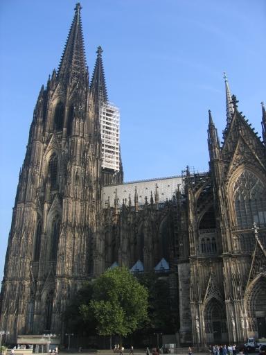
I reached Cologne, the day before the meeting started. The journey was smooth and in addition, the weather in Germany was cool and pleasant. Just after getting off at Hubbernhof (Cologne rail station), the sight of tall and picturesque Cologne Cathedral energized me from travel weariness. The ensuing break was good for recuperation until the start of meeting on the next day.

On the initial day, after registration, a considerable search was made to locate the assigned area for setting up of poster as they were not serially arranged. The keynote presentation on the first day titled “Stem cells for CNS Repair” given by Anders Bjorklund, Lund University, Sweden, identified the development of stem cells to treat Parkinson’s disease (PD). The use of primary neuroblasts obtained from aborted fetuses was successfully demonstrated in clinical trials of PD. A study using transplants of transgenic mice expressing GFP under control of rat tyrosine hydroxylase promoter into striatum of neonatal rats having dopaminergic neuronal lesion was presented. However, it was mentioned that immunogenicity was one of the major drawbacks for successful long-term effect. The group is currently trying to develop dopaminergic neuroblasts from neural progenitors and embryonic stem cells for cellular therapy. The development of molecular imaging probes to monitor such a therapy was also addressed.

From next day, the lectures were organized as plenary sessions at mornings and Concurrent Symposium sessions at noon. In the first session, Heiss WD discussed about primary role of 11C-Flumazenil PET in evaluation of malignant brain edema following focal cerebral ischemia. A presentation by Alnawaz Rehemtulla gave a broad overview of understanding different mechanisms of cancer biology through molecular imaging. Some of them included p53 gene activity imaging via luciferase reporter in a transgenic animal model, imaging apoptosis using silencing domains and imaging Akt activity in tumors. A talk by Helmut Maecke focused on pharmacokinetics and biodistribution characteristics of radiometal labeled peptides (111In-DOTATOC, 177Lu-DOTATOC) on somatostatin and Substance P receptors in imaging of tumors. In novel reporter gene session, a pro-drug approach for restoration of dopamine function in PD was presented by Eberling J. A PET substrate [18F]fluoro-L-m-tyrosine (FMT) was used as a reporter probe to measure Aromatic L-amino acid decarboxylase (AADC) activity in striatum of MPTP induced PD monkey models. L-dopa was used as a pro-drug to restore dopaminergic activity in those monkey models. AADC is a key enzyme in final conversion of L-dopa to dopamine. AADC gene was inserted into an adeno-associated virus (AAV) vector, introduced directly into striatum and FMT-PET imaging was performed to determine AADC transgene expression and activity. A constitutive expression for 45 months post-treatment, as observed by FMT imaging and post-mortem immunohistological studies were of particular interest. A preliminary clinical trial in a PD patient with a moderate success was also presented. A concept of artificial reporter gene for MRI using Chemical Exchange Saturation Transfer (CEST) was introduced by Gilad of Johns Hopkins School of Medicine. It basically involved using an oligonucleotide sequence capable of generating CEST polypeptide agents which can produce contrast based on chemical exchange rate of the agent protons with water. A gene coding for polylysine (a stretch of 200 amino acids) was quoted as an example of such a reporter sequence.

The multimodality imaging talks mostly focused on combined PET-MR, discussing about various benefits over PET-CT and instrumentation criterions to be fulfilled for its success.

Poster sessions were in the evenings and categorized over three days. There were more than 600 poster presentations on varied aspects of molecular imaging. Perhaps for that reason, the time limit for each presentation was just 2 minutes, in which hardly one could elucidate their work or comprehend that of others. I had few queries on my poster regarding the intended mode of adenoviral delivery in animal models, the type of imaging and so on.

Overall, it was a tremendous learning experience for a first time attendee like me. I hope to gain more knowledge in future meetings.

Barring the meeting, the city of Cologne was splendid with its rich blend of traditional and modern culture. The ascent to the top of Cologne Cathedral (Tower height of 157m and world’s tallest structure until 1884!!!) and viewing the city through miniature windows at multiple angles was amusing. The central shopping streets were always buzzing with charming people and open-air cafes. The dinner cruise on the river Rhine and the concert at Gurzenich are few of the memories to cherish for a long time to come…

Tuesday, December 14, 2004

Third International Workshop on Biomedical Imaging-Fukui 2004

Hi folks

This is my first blog post. Just finished exciting talks on Molecular Imaging given by top nuclear scientists such as Dr H. Wagner Jr, Dr MJ welch, Dr Barry Seigal, Dr Minoshima and others. The presentations of their topics were interesting as well as informative about the current trends and prospects occuring in this field.
Of particular interest was Dr Wagner's talk on "new definition of diesease" wherein he proposed the idea of future Human chip supposedly containing a patient's diesease profile (text or images)including his genome, with which a Physician can get to know the present and future status of a patient after feeding it to a computer. However the creation or validation of the data and its implementation is of a big concern unless some potential IT majors such a Google or Microsoft helps in developing such a chip.