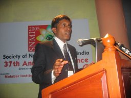
I attended the 37th Annual Conference of Society of Nuclear Medicine, India (SNMI) 2005 held at Calicut in the state of Kerala, India from 7th -11th December, 2005. It was the first time for me to attend an International conference related to Nuclear medicine held in India.
The conference venue was at Kadavu Resort, located in a sprawling nine-acre plot along the side of the backwaters, with groves of swaying, graceful coconut palms facing the dancing ripples on the water.
The theme of the conference was chosen to be “Scroll into the era of Fusion Imaging” to reflect the rapid changes and the future opportunities of Nuclear Medicine in India with a global perspective.
The Scientific Programme consisted of Keynote addresses, plenary lectures and Specialty sessions delivered by pioneers and experts of Nuclear medicine and molecular imaging from India and abroad. In addition, symposia, panel discussion and free paper sessions were also part of the programme. There were nearly 250 delegates including students, with experts coming from USA, UK, Singapore, Hong Kong and Korea. I was the only representative from Japan. There were 35 poster presentations and 40 oral presentations in free paper session covering various clinical and basic research topics of Nuclear medicine.
The welcome address was given by Dr. P.S. Soni, Head, PET Cyclotron Division, Radiation Medicine Centre, Mumbai, India. The initial day’s lectures had two sessions - Fusion Imaging (Technical and Clinical) and Gastroenterology coupled with symposium on “sentinel node biopsy” and a panel discussion on “Interstitial lung disease”. The fusion imaging lectures covered basics and radiation safety of SPECT-CT and PET-CT fusion imaging. The significant factors contributing to attenuation correction accuracy of fused images such as CT acquisition time, motion artifacts, slice thickness variations and reconstruction strategies were mentioned. There were also talks on clinical usage of SPECT-CT in skeletal, infectious and oncological disorders. Few clinical cases of implant infections and pyrexia of unknown origin (PUO) were imaged with 99mTc-labelled leukocyte or Gallium SPECT-CT and accurate localization and quantification of the infection was reported. In the Gastroenterology session, talks were given on the relative challenges of ERCP and MRCP in hepatobiliary scintigraphy. Advances in the usage of radioisotope liver scan for diffuse hepatic disease was also presented in the same session.
In the next day among the special orations, Homi Bhaba oration was given by Dr Joseph Mantil, who gave a general talk of tools of Molecular Imaging currently available and stressed on importance of early diagnosis of atherosclerosis using FDG imaging. In the cardiology session, benefits of myocardial perfusion scan were discussed. The imaging techniques other than conventional coronary angiography in patient management such as quantifying atheroma burden using Multislice CT coronary calcium scoring or EBCT, use of perfusion scan as prognostic indicator of cardiac dysfunction, use of perfusion MR imaging to localize subendocardial hypoperfusion were presented. Among the free paper session, few lectures were of notable interest. Most of them were either clinical studies or evaluation of cyclotron or SPECT-CT imaging applications. An indigenous user friendly and easy to use Quality control test kit for 99mTc-radiopharmaceuticals called QTECH was introduced. My talk was scheduled in the post-lunch session and as expected it gathered little audience after heavy meals! Though there were not many queries immediately after my talk, a few came up during tea break.
The next day’s session was entirely devoted to applications of PET-CT in different clinical disorders such as Lymphoma, breast cancer, thyroid cancer and other benign disorders. Here I would like to mention that currently there are only two functional cyclotrons in the country – one at Mumbai (BARC) and other at Hyderabad (Apollo Hospital). There is only one dedicated PET camera at Tata Memorial Hospital and three coincidence gamma PET cameras at Hinduja Hospital, Jaslok Hospital and Bombay Hospital all being in Mumbai. At Hyderabad, a PET-CT machine was installed this June and has started functioning. Squarely speaking, this is a negligible contribution to a country having a population of more than a billion and they too are recent installations. The major reasons for this include cost-effectiveness, government restrictions and low awareness among the health care planners and medical fraternity outside imaging community. But with increasing pressure from imaging societies and multinational companies, over the coming 2-3 years, plans have been charted to install few more cyclotrons and PET-CT in Delhi, Kolkata and Bangalore. The need for them was well appraised with leading nuclear medicine specialists giving talks on forthcoming clinical applications of PET-CT.
The final day was concluded with plenary lecture from Dr. June Key Chung on molecular genetic imaging using Nuclear medicine methods and an oration from Dr P.S.Soni.
Apart from the brainstorming academic sessions, the organizers had staged an exquisite cultural extravaganza of the state of Kerala. So our evenings were occupied with local art shows, music shows and cruise to a nearby island. Overall, I had a first hand learning experience of present standards of Nuclear medicine in India. Though the emphasis at present was on clinical aspects, a fair amount of research seems likely to begin henceforth. As a special note, I was pleased to know the SNMI President, Dr. Sanjay Gambhir and Dr P.S. Soni had part of their research careers in Japan and was more surprised to hear Japanese from them!

No comments:
Post a Comment